


Home / Training / Manuals / Atlas of breast cancer early detection / Cases
Atlas of breast cancer early detection
Go back to the list of case studies
.png) Click on the pictures to magnify and display the legends
Click on the pictures to magnify and display the legends
| Case number: | 186 |
| Age: | 74 |
| Clinical presentation: | Postmenopausal woman with average risk of developing breast cancer presented with a right breast lump noticed more than 2 years ago. She now also had right nipple retraction. |
 |
Mammography:
 |  |
 | 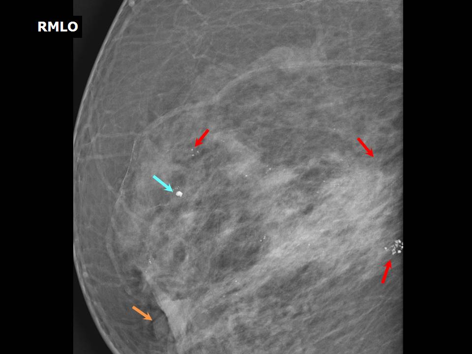 |
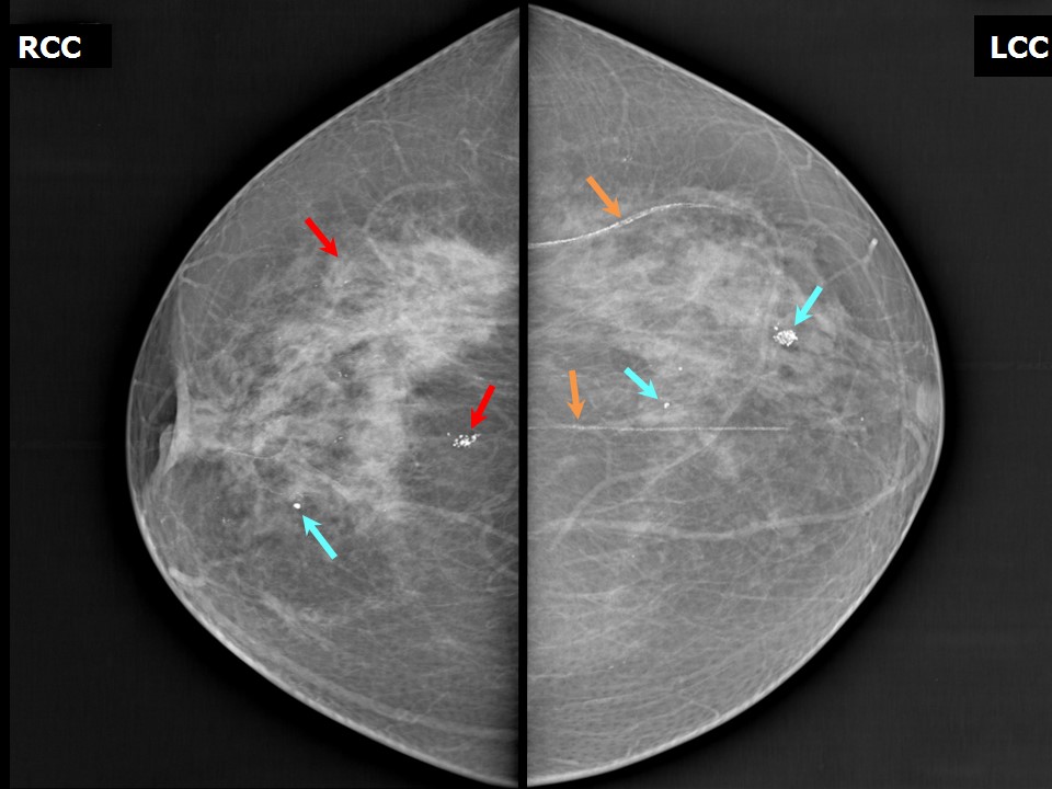
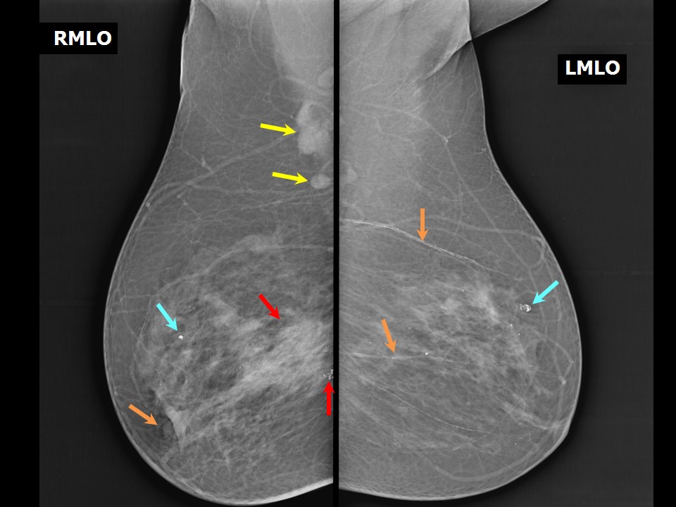
| Breast composition: | ACR category b (there are scattered areas of fibroglandular density) | Mammography features: |
| ‣ Location of the lesion: | Right breast, upper outer quadrant at 910 oclock, anterior, middle, and posterior thirds |
| ‣ Mass: | |
| • Number: | 0 |
| • Size: | None |
| • Shape: | None |
| • Margins: | None |
| • Density: | None |
| ‣ Calcifications: | |
| • Typically benign: | None |
| • Suspicious: | Fine microcalcifications and clustered pleomorphic microcalcifications |
| • Distribution: | Segmental, grouped |
| ‣ Architectural distortion: | None |
| ‣ Asymmetry: | Focal |
| ‣ Intramammary node: | None |
| ‣ Skin lesion: | None |
| ‣ Solitary dilated duct: | None |
| ‣ Associated features: | Enlarged axillary nodes, focal asymmetry, trabecular thickening, fine microcalcifications, and clustered pleomorphic microcalcifications |
| Breast composition: | ACR category b (there are scattered areas of fibroglandular density) | Mammography features: |
| ‣ Location of the lesion: | Right breast, upper inner quadrant at 1 oclock, middle third |
| ‣ Mass: | |
| • Number: | 0 |
| • Size: | None |
| • Shape: | None |
| • Margins: | None |
| • Density: | None |
| ‣ Calcifications: | |
| • Typically benign: | Yes, round |
| • Suspicious: | None |
| • Distribution: | Diffuse |
| ‣ Architectural distortion: | None |
| ‣ Asymmetry: | None |
| ‣ Intramammary node: | None |
| ‣ Skin lesion: | None |
| ‣ Solitary dilated duct: | None |
| ‣ Associated features: | None |
| Breast composition: | ACR category b (there are scattered areas of fibroglandular density) | Mammography features: |
| ‣ Location of the lesion: | Left breast, upper outer quadrant at 1 oclock, anterior and middle thirds |
| ‣ Mass: | |
| • Number: | 0 |
| • Size: | None |
| • Shape: | None |
| • Margins: | None |
| • Density: | None |
| ‣ Calcifications: | |
| • Typically benign: | Yes, round, coarse heterogeneous, and vascular calcifications |
| • Suspicious: | None |
| • Distribution: | Diffuse |
| ‣ Architectural distortion: | None |
| ‣ Asymmetry: | None |
| ‣ Intramammary node: | None |
| ‣ Skin lesion: | None |
| ‣ Solitary dilated duct: | None |
| ‣ Associated features: | None |
Ultrasound:
 |  |
 | 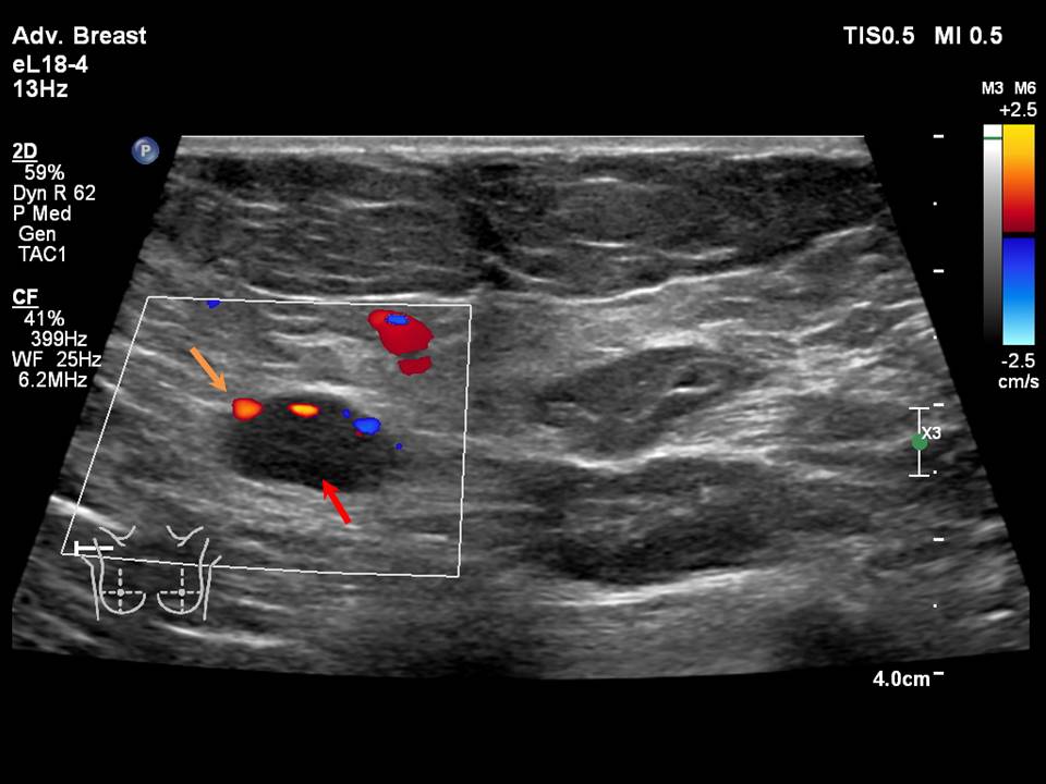 |
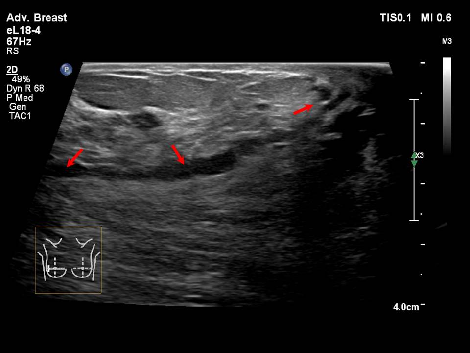
| Ultrasound features: None | |
| ‣ Mass | |
| • Location: | None |
| • Number: | 0 |
| • Size: | None |
| • Shape: | None |
| • Orientation: | None |
| • Margins: | None |
| • Echo pattern: | None |
| • Posterior features: | No posterior features |
| ‣ Calcifications: | Intraductal microcalcifications |
| ‣ Associated features: | Focal asymmetry, dilated ducts, axillary dysmorphic lymphadenopathy with non-hilar vascularity on colour flow mapping |
| ‣ Special cases: | None |
BI-RADS:
BI-RADS Category: 5 (highly suggestive of malignancy)Further assessment:
Further assessment advised: Referral for cytologyCytology:
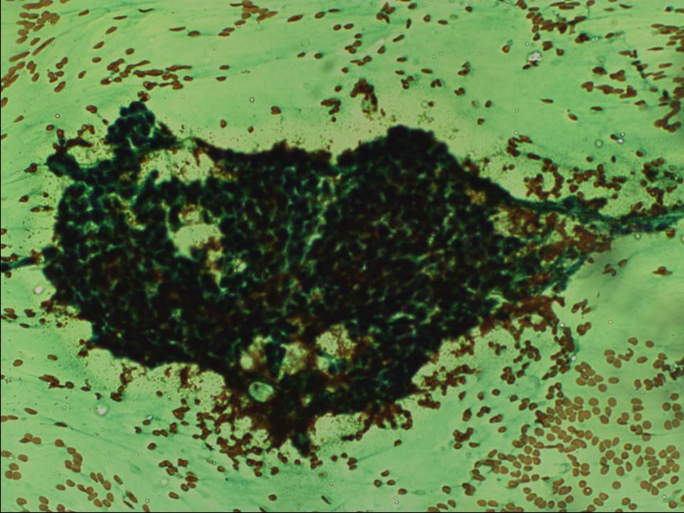 | 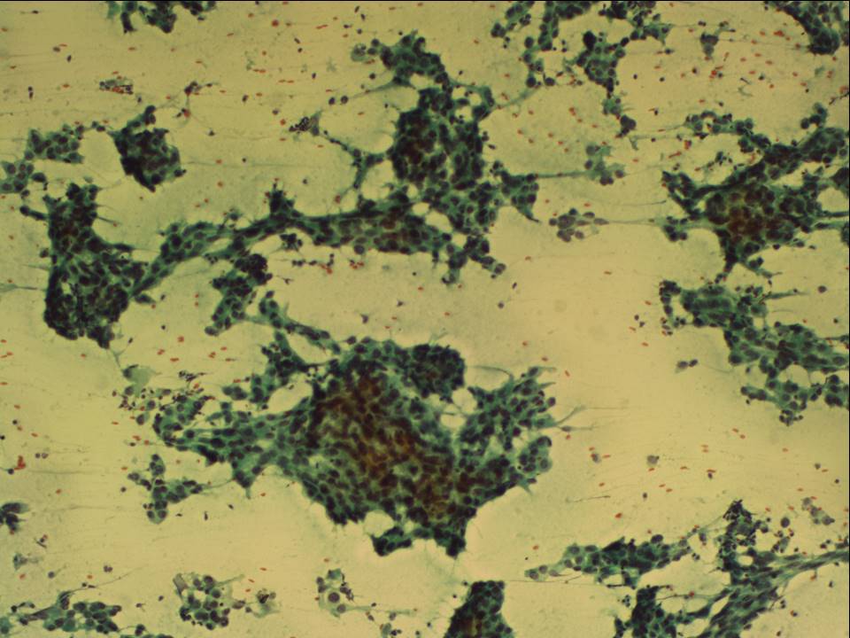 |
| Cytology features: | |
| ‣ Type of sample: | FNAC |
| ‣ Site of biopsy: | |
| • Laterality: | Right |
| • Quadrant: | Upper half lumpish feel on palpation |
| • Localization technique: | Palpation |
| • Nature of aspirate: | Whitish |
| ‣ Cytological description: | Smears from first pass show proteinaceous fluid, foamy macrophages, and a few cohesive benign ductal epithelial cells. Smears from second pass show loosely cohesive sheets of ductal epithelial cells with nuclear pleomorphism, high N:C ratio, and hyperchromasia. Myoepithelial cells are absent |
| ‣ Reporting category: | Suspicious, probably in situ or invasive carcinoma |
| ‣ Diagnosis: | Suspicious for malignancy, probably in situ or invasive carcinoma |
| ‣ Comments: | None |
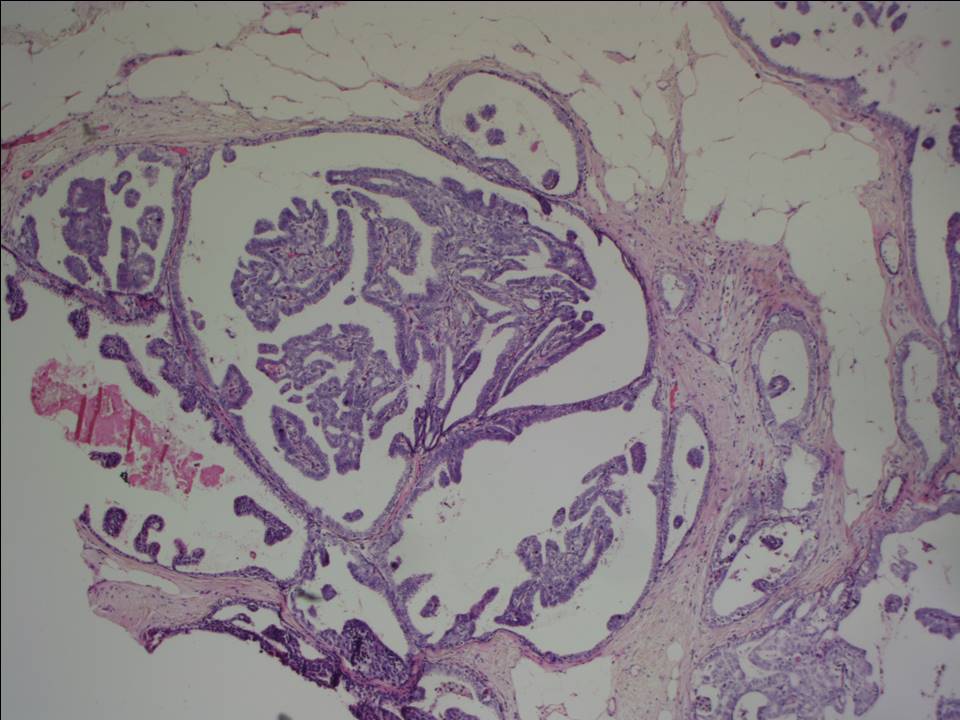
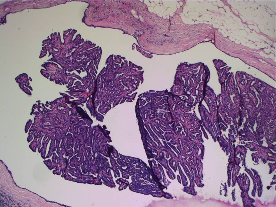
Histopathology:
MRM
 |  |
 |  |
 |
| Histopathology features: | |
| ‣ Specimen type: | MRM |
| ‣ Laterality: | Right |
| ‣ Macroscopy: | Right MRM specimen (27.5 × 12.5 × 3.5 cm) with skin flap (21.0 × 7.5 cm). The nipple is retracted. On serial sectioning, multiple firm greyish white areas (7.0 × 3.0 × 3.0 cm, 3.0 × 2.0 × 2.5 cm, and 2.5 × 2.5 × 2.0 cm) are seen over the upper half of the breast parenchyma. Distance from base (posterior margin of resection) is 2 cm. Largest node (2.5 × 1.5 × 1.5 cm) was dissected. The cut surface is whitish |
| ‣ Histological type: | Invasive breast carcinoma of no special type |
| ‣ Histological grade: | Grade 2 (3 + 2 + 2 = 7) |
| ‣ Mitosis: | 12 |
| ‣ Maximum invasive tumour size: | 5.5 cm (other tumours 3 cm and 2.6 cm in greatest dimension) |
| ‣ Lymph node status: | 5/20 |
| ‣ Peritumoural lymphovascular invasion: | Present |
| ‣ DCIS/EIC: | DCIS: cribriform, solid, papillary, and micropapillary type, moderate nuclear grade mainly, no necrosis. DCIS also involves the large lactiferous ducts. Paget disease of nipple present. EIC present |
| ‣ Margins: | Free of tumour |
| ‣ Pathological stage: | pT3(3)N2 |
| ‣ Biomarkers: | ER positive, PR positive, and HER2 negative |
| ‣ Comments: | The adjacent breast shows fibrocystic change, apocrine metaplasia, papilloma, and multiple areas with UDH |
Case summary:
| Postmenopausal woman with average risk of developing breast cancer presented with right breast lump noticed more than 2 years ago. Now with right nipple retraction also. Diagnosed as right breast outer quadrant focal asymmetry with calcifications of suspicious morphology, BI-RADS 5 (highly suggestive of malignancy) on imaging, as suspicious for malignancy on cytology, and as invasive breast carcinoma of no special type with EIC pT3(3)N2 with Paget disease on histopathology. |
Learning points:
|